| Abbildung |
|---|
| |
|
|
MR-Angiographie
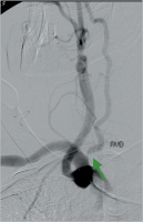
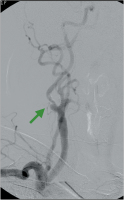
Abbildung 1: Präoperative MR-Angiographie: Verschluss des Truncus brachiocephalicus
(grüner Pfeil) und der linken Art. carotis communis und interna, hochgradige
Abgangsstenose der linken Art. vertebralis (grüner Pfeil).
Keywords: Arteria carotis,
Gefäßmedizin,
MR-Angiographie
|
| |
| |
|
|
Angiographie
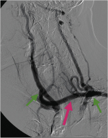
Abbildung 2: Postoperative Angiographie: Bypass (roter Pfeil) von der linken Art.
subclavia (peripher des Abgangs der Art. vertebralis, grüner Pfeil) auf die rechte Art.
carotis communis (grüner Pfeil).
Keywords: Angiographie,
Arteria carotis,
Gefäßmedizin
|
| |
| |
|
|
Atherosklerose
Abbildung 3: Atherosklerotisches Art. carotis-interna-Aneurysma links (grüner Pfeil).
Keywords: Arteria carotis,
Gefäßmedizin,
Operationssitus
|
| |
| |
|
|
PTFE-Interponat
Abbildung 4: Nach Resektion und 6-mm-PTFE-Interponat (weißer Pfeil).
Keywords: Gefäßmedizin,
Operationssitus,
PTFE-Interponat
|
| |
| |
|
|
Arteria carotis interna
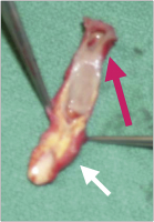
Abbildung 5: Resezierte und eröffnete
Art. carotis interna; das Aneurysma
(roter Pfeil) ist eröffnet, proximal im
Karotisbulbus atherosklerotische Stenose
(weißer Pfeil).
Keywords: Arteria carotis,
Gefäßmedizin,
Operationssitus
|
| |
| |
|
|
CT-Angiographie
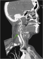
Abbildung 6: CT-Angiographie der rechten Karotis bei Zustand nach Thrombendarterektomie
und Patchverschluss vor 14 Jahren und asymptomatischer Restenose
(grüner Pfeil).
Keywords: Arteria carotis,
CT-Angiographie,
Gefäßmedizin
|
| |
| |
|
|
CT-Angiographie
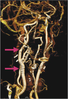
Abbildung 7: Postoperative CT-Angiographie nach 6-mm-PTFE-Interponat von der Art. carotis communis auf die Art. carotis interna (rote Pfeile).
Keywords: Arteria carotis,
CT-Angiographie,
Gefäßmedizin
|
| |
| |
|
|
Arteria carotis
Abbildung 8: Resezierte Art. carotis interna; die Nahtreihe des alten Patch ist mit dem roten Pfeil markiert.
Keywords: Arteria carotis,
Gefäßmedizin,
Operationssitus
|
| |
| |
|
|
Arteria carotis
Abbildung 9: Luminale Ansicht der resezierten Art. carotis interna; die Pinzette markiert
das bulbusnahe, atheromatöse Areal der Art. carotis communis/interna, der rote
Pfeil markiert die Nahtreihe des alten Patch.
Keywords: Arteria carotis,
Gefäßmedizin,
Operationssitus
|
| |
| |
|
|
Subtraktionsangiographie
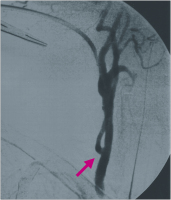
Abbildung 10: Subtraktionsangiographie. Hochgradige Abgangsstenose der rechten
Art. vertebralis.
Keywords: Arteria vertebralis,
Gefäßmedizin,
Subtraktionsangiographie
|
| |
| |
|
|
Subtraktionsangiographie
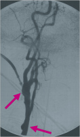
Abbildung 11: Subtraktionsangiographie. Hochgradige, exzentrische Abgangsstenose
der rechten Art. carotis interna (grüner Pfeil).
Keywords: Arteria carotis,
Gefäßmedizin,
Subtraktionsangiographie
|
| |
| |
|
|
Kontrollangiographie
Abbildung 12: Intraoperative Kontrollangiographie nach simultaner Eversionsendarterektomie
der rechten Art. carotis interna und Transposition der rechten Art. vertebralis
in die rechte Art. carotis communis (roter Pfeil).
Keywords: Angiographie,
Arteria carotis,
Gefäßmedizin
|
| |
| |
|
|
Kontrollangiographie
Abbildung 13: Intraoperative Kontrollangiographie in einer 2. Ebene nach simultaner
Eversionsendarterektomie der rechten Art. carotis interna (roter Pfeil) und Transposition
der rechten Art. vertebralis in die rechte Art. carotis communis (roter Pfeil).
Keywords: Angiographie,
Arteria carotis,
Gefäßmedizin
|
| |
| |
|
|
Ascendensbypass
Abbildung 14: Ascendensbypass (weißer Pfeil) auf Truncus brachiocephalicus und
Art. carotis communis links mit angeschlossenem Conduit (roter Pfeil; grüner Pfeil
Aorta ascendens).
Keywords: Arteria carotis,
Ascendensbypass,
Gefäßmedizin
|
| |
| |
|
|
Abschlussangiographie
Abbildung 15: Intraoperative Abschlussangiographie nach Stenteinbringung (Conduit
über den der thorakale Stentgraft retrograd eingeführt wurde, grüner Pfeil; Subclaviatransposition
roter Pfeil; Bypass auf Truncus brachiocephalicus und Art. carotis
communis links, weißer Pfeil).
Keywords: Angiographie,
Gefäßmedizin,
Stent
|
| |
| |
|
|
Arteria carotis
Abbildung 16: Elongierte Art. carotis interna nach Stentangioplastie mit symptomatischer
Knickstenose (weißer Pfeil).
Keywords: Arteria carotis,
Gefäßmedizin,
Stent
|
| |
| |
|
|
Arteria carotis
Abbildung 17: Karotis nach Eversionendarterektomie und Kürzung; der weiße Pfeil
markiert die Lokalisation der alten Knickstenose.
Keywords: Arteria carotis,
Eversionendarterektomie,
Gefäßmedizin
|
| |
| |
|
|
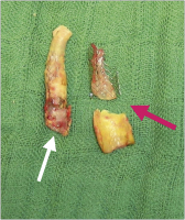
Arteria carotis
Abbildung 18: Evertierter Stent aus der Art. carotis interna (weißer Pfeil) und Art.
carotis communis (roter Pfeil).
Keywords: Arteria carotis,
Gefäßmedizin,
Stent
|
| |
| |
 Suche Medizinische Publikationen
 Suche Abbildungen und Grafiken
 Medizinprodukte - Labortechnik
 Antidotarium (Stand 2012)
 Phytokodex (Stand 2012)
 Fachbücher
 Patientenliteratur