Suche Medizinische Publikationen
 Suche Abbildungen und Grafiken
 Medizinprodukte - Labortechnik
 Antidotarium (Stand 2012)
 Phytokodex (Stand 2012)
 Fachbücher
 Patientenliteratur
| Bellmann B et al. | ||||||||||||||||||||||
|---|---|---|---|---|---|---|---|---|---|---|---|---|---|---|---|---|---|---|---|---|---|---|
|
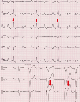
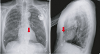
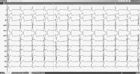
Case Report: Pacemaker Mimics Ventricular Extrasystoles?
Journal für Kardiologie - Austrian Journal of Cardiology 2017; 24 (11-12): 286-287 Volltext (PDF) Abbildungen
|
||||||||||||||||||||||

Verlag für Medizin und Wirtschaft |
|
||||||||
|
Abbildungen und Graphiken
|
|||||||||
| copyright © 2000–2026 Krause & Pachernegg GmbH | Sitemap | Datenschutz | Impressum | |||||||||
|
|||||||||