Suche Medizinische Publikationen
 Suche Abbildungen und Grafiken
 Medizinprodukte - Labortechnik
 Antidotarium (Stand 2012)
 Phytokodex (Stand 2012)
 Fachbücher
 Patientenliteratur
| Schmidinger S et al. | ||||||||||||||||
|---|---|---|---|---|---|---|---|---|---|---|---|---|---|---|---|---|
|
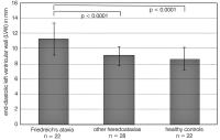
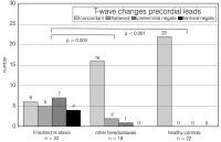
Cardiomyopathy in patients with Friedreich's ataxia - appearance and diagnostic value
Journal of Clinical and Basic Cardiology 2000; 3 (3): 167-171 PDF Summary Figures
|
||||||||||||||||

Verlag für Medizin und Wirtschaft |
|
||||||||
|
Figures and Graphics
|
|||||||||
| copyright © 2000–2026 Krause & Pachernegg GmbH | Sitemap | Datenschutz | Impressum | |||||||||
|
|||||||||