| Binder T |
|---|
Three-Dimensional Echocardiography - Principles and Promises
Journal of Clinical and Basic Cardiology 2002; 5 (2): 149-152
PDF Summary Figures
| Figure |
|---|
| |
|
|
3D-Echokardiographie
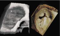
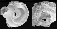
Figure 1: Three-dimensional reconstruction of the mitral valve (left) as seen from the left atrium. An anatomical specimen for comparison is shown on the right.
Keywords: 3D,
3D,
echocardiography,
Echokardiographie,
Mitral valve,
Mitralklappe,
Reconstruction,
Rekonstruktion
|
| |
| |
|
|
3D-Echokardiographie
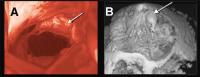
Figure 2: Patient with a partial flail leaflet as seen during mitral valve repair. 3D-Echo is able to demonstrate the location and morphology on the defect (arrow).
Keywords: 3D,
3D,
echocardiography,
Echokardiographie,
Mitral valve,
Mitralklappe,
Reconstruction,
Rekonstruktion
|
| |
| |
|
|
3D-Echokardiographie
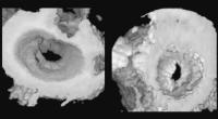
Figure 3: Three-dimensional reconstruction of a patient with mitral stenosis, morphologic features such as doming of the anterior leaflet and commissural fusion can be noticed. In addition, quantification of mitral valve area can be performed.
Keywords: 3D,
3D,
echocardiography,
Echokardiographie,
Mitral valve,
Mitralklappe,
Stenose,
Stenosis
|
| |
| |
|
|
3D-Echokardiographie
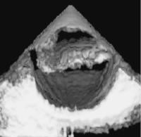
Figure 4: Dissection of the descending aorta. The true lumen is separated from the false lumen by an intima flap.
Keywords: 3D,
3D,
Aorta descendens,
Aorta descendens,
dissection,
Dissektion,
echocardiography,
Echokardiographie,
Intima,
Intima
|
| |
| |
|
|
3D-Echokardiographie
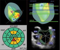
Figure 5: Three-dimensional analysis of left ventricular function allows computation of global and regional ejection fraction (bottom) and the three-dimensional display of the ventricle (top panel).
Keywords: 3D,
3D,
Auswurffraktion,
echocardiography,
Echokardiographie,
Ejection fraction,
Ejektion,
Function,
Funktion,
ventricle,
Ventrikel
|
| |
| |
|
|
3D-Echokardiographie
Figure 6: Model of the mitral valve (in a patient with mitral stenosis) created from a 3-dimensional dataset using stereolithography.
Keywords: 3D,
3D,
Echokardiographie,
Echokardiographie,
Mitral valve,
Mitralklappe,
Stenose,
Stenosis,
Stereolithographie,
Stereolithography
|
| |
| |
|
|
 Suche Medizinische Publikationen
 Suche Abbildungen und Grafiken
 Medizinprodukte - Labortechnik
 Antidotarium (Stand 2012)
 Phytokodex (Stand 2012)
 Fachbücher
 Patientenliteratur